Why hypertension can cause a stroke and when surgery saves lives
Hypertensive stroke with cerebral hemorrhage is one of the most dangerous conditions that can occur suddenly. Imagine: a small blood vessel deep within the brain ruptures, and blood spills directly into the brain tissue, forming a hematoma (blood clot). This compresses nerve cells, disrupts blood flow, and triggers a chain reaction of damage.
Such hypertensive intracerebral hematomas account for 10-15% of all strokes, but they account for up to 40% of deaths from this disease. Mortality rates reach 40-70%, and among survivors, up to 75% are left with severe disabilities. The main culprit is chronic, uncontrolled hypertension.
Why is this happening?
Over the course of many years, high blood pressure wears down the walls of small arteries in the brain (especially in the deep structures: the basal ganglia, thalamus, cerebellum, and pons). The walls become brittle, like an old rubber tube. Lipohyalinosis (fat and protein deposits) develops, and tiny bulges called Charcot-Bouchard microaneurysms appear. A sudden surge in pressure can cause the vessel to rupture.
Blood doesn't just take up space—it's toxic to the brain. Its breakdown products cause inflammation, swelling, and death of adjacent neurons. A hematoma mechanically compresses tissue, displaces brain structures, and can rupture into the ventricles, further worsening the situation.
How to recognize and quickly diagnose
Symptoms develop rapidly (unlike ischemic stroke, where there is sometimes a “window”):
A sudden, severe headache (“like never before in my life”).
Vomiting, impaired consciousness (from drowsiness to coma).
Weakness in half of the body (hemiparesis), problems with speech, coordination or vision.
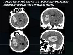
The gold standard for diagnosis is computed tomography (CT) of the head. It takes minutes, clearly shows the hematoma as a bright white spot, measures its volume (often using a simple ABC/2 formula), and detects swelling and displacement of brain structures. If necessary, CT angiography is performed to rule out other causes (aneurysms, malformations) or to detect a "spot sign"—an indication of ongoing bleeding.
MRI is useful later to clarify details, but in the acute stage, CT is faster and more accessible.
Treatment: surgery or conservative therapy
Most patients (70–85%) receive conservative treatment: strict blood pressure control (not too abruptly, so as not to impair cerebral blood flow), swelling control (mannitol, etc.), respiratory support, and complication prevention. This is the basis for everyone.
Surgical hematoma removal is performed selectively—in approximately 10–30% of patients, depending on the region and the clinic's capabilities. Surgery is not a cure-all and is unnecessary for small hematomas (<30 ml) or in the most severe cases (deep coma with irreversible brainstem damage).
When is surgery most often considered?
Cerebellar hematomas with a volume of >10–15 ml (or a diameter of >3 cm) – here the risk of rapid compression of the brainstem is very high, and surgery is often life-saving.
Large superficial (lobar) hematomas >30 ml with deterioration of consciousness and mass effect.
Signs of increasing edema, displacement of structures or breakthrough into the ventricles (then a drain is placed to remove cerebrospinal fluid).
Operation options:
Open craniotomy is the classic wide approach. It allows for complete removal of clots, but is traumatic, especially for deep hematomas.
Minimally invasive methods (preferred today):
Stereotactic puncture with blood aspiration and administration of clot-dissolving drugs (for example, according to the MISTIE protocol).
Endoscopic removal: an endoscope is inserted through a small opening, the doctor sees everything on the screen and carefully evacuates the hematoma.
External ventricular drainage - for hydrocephalus.
Surgery is considered only when the potential benefit outweighs the risks of the surgical trauma itself.
What Science Says About Effectiveness
Classic studies (STICH I and II) have shown that open surgery generally offers little advantage over high-quality conservative therapy for most patients with deep hematomas. However, for lobar (superficial) hematomas and cerebellar hemorrhages, the benefit is more significant.
An important recent study, ENRICH (2024), confirmed that early minimally invasive hematoma evacuation improves functional outcomes (ability to live independently) at 6 months, especially for lobar hemorrhages of 30–80 ml. For deep hemorrhages (in the basal ganglia), the effect is less pronounced or absent. Mortality was lower in the surgical group (9.3% versus 18% in the control group).
Overall, surgery helps strictly selected patients: young, with moderately severe conditions, a large superficial hematoma, and signs of deterioration. The goal is not just survival, but maintaining quality of life.
Forecast: Harsh Reality and Chances for Recovery
Hemorrhagic stroke remains one of the most severe vascular events:
30-day case fatality rate: 35–50% (half of deaths occur in the first 48 hours);
1-year mortality: up to 50–70%;
Only 20–30% of survivors return to independent living.
But the figures vary depending on the tactics used. Modern meta-analyses and studies (STICH, MISTIE III, ENRICH) show:
Surgery is not a panacea for everyone, but in a properly selected group it reduces overall mortality by ~9% and increases the chance of a good functional outcome by ~5%.
In case of cerebellar hematomas, surgery reduces mortality by 30–40%.
For medium and large superficial hematomas, minimally invasive removal within the first 24 hours significantly improves the prognosis.
Key factors worsening outcome: large hematoma volume, low level of consciousness on admission, blood breakthrough into the ventricles, age >80 years, positive “spot sign” and comorbidities.
Conclusion: The best operation is the one that is not needed.
Neurosurgery for hypertensive stroke has evolved from the dogma of "operating on everyone" to personalized medicine. Today, it's not a choice between "cutting" and "waiting," but rather precise adjustments: the location of the clot, its volume, how the brain responds, what technologies are available in the clinic, and whether the patient can undergo rehabilitation.
But the main tool in the fight against this pathology lies in the hands of each of us. Monitoring blood pressure, regularly taking antihypertensive medications, quitting smoking and alcohol abuse, and being physically active are not just abstract recommendations; they are real protections for the cerebral blood vessels.
Hemorrhagic stroke develops rapidly, but it can be prevented. And if disaster does occur, modern medicine offers hope: timely diagnosis, competent intensive care, and, when strictly indicated, minimally invasive surgery can turn a fatal scenario into a recovery.
Why hypertension can cause a stroke and when surgery saves lives